Staining: A Detailed Overview

Basophilic Staining
Definition
Basophilic refers to the uptake of basic (alkaline) dyes by certain cellular components. The term “basophilic” comes from “base-loving,” indicating that these structures have an affinity for basic dyes.
Chemical Basis
Basophilic structures are typically rich in acidic components, such as nucleic acids (DNA and RNA) and certain proteins. These acidic substances attract and bind basic dyes, such as hematoxylin.
Common Basophilic Dyes
- Hematoxylin: One of the most commonly used basic dyes. It stains nucleic acids and rough endoplasmic reticulum blue to purple.
- Toluidine Blue: Another basic dye that stains acidic cell components blue.
Basophilic Structures
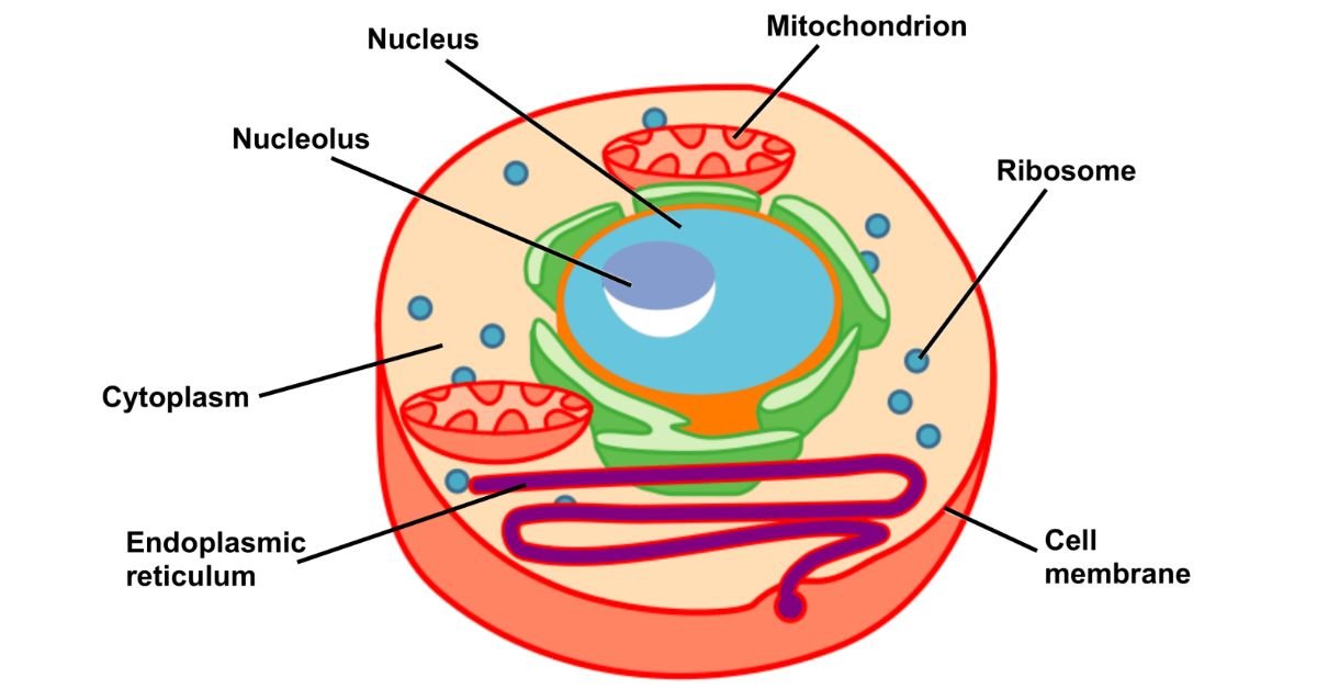
- Nucleus: Contains DNA and RNA, which bind strongly to basic dyes.
- Rough Endoplasmic Reticulum: Contains RNA in ribosomes, which also bind to basic dyes.
- Cartilage Matrix: Contains acidic polysaccharides that attract basic dyes.
Acidophilic Staining
Definition
Acidophilic refers to the uptake of acidic dyes by certain cellular components. The term “acidophilic” comes from “acid-loving,” indicating that these structures have an affinity for acidic dyes.
Chemical Basis
Acidophilic structures are typically rich in basic components, such as cytoplasmic proteins and certain extracellular matrix proteins. These basic substances attract and bind acidic dyes, such as eosin.
Common Acidophilic Dyes
- Eosin: A widely used acidic dye that stains cytoplasmic proteins and extracellular matrix proteins pink to red.
- Orange G: Another acidic dye that can be used to stain proteins orange.
Acidophilic Structures
- Cytoplasm: Contains many basic proteins that bind to acidic dyes.
- Collagen Fibers: Part of the extracellular matrix, these proteins also attract acidic dyes.
- Muscle Fibers: Rich in proteins that bind acidic dyes.
Practical Applications
Hematoxylin and Eosin (H&E) Staining
H&E is the most common histological staining technique, utilizing both basophilic and acidophilic dyes. Hematoxylin stains the nuclei blue, while eosin stains the cytoplasm and extracellular matrix pink. This contrast allows for detailed visualization of tissue architecture and cellular morphology.
Steps of H&E
- Fixation: Preserving the tissue sample to prevent degradation.
- Embedding: Encasing the tissue in a solid medium, usually paraffin, for sectioning.
- Sectioning: Cutting thin slices of the tissue for mounting on slides.
- Staining with Hematoxylin: Staining the nuclei and other basophilic structures.
- Differentiation: Removing excess hematoxylin to improve contrast.
- Staining with Eosin: Staining the cytoplasm and other acidophilic structures.
- Mounting: Covering the stained tissue with a coverslip for microscopy.
Special Stains
In addition to H&E, other techniques target specific cell components:
- Masson’s Trichrome: Differentiates muscle, collagen, and fibrin using three dyes.
- Periodic Acid-Schiff (PAS): Stains carbohydrates, such as glycogen and mucins, magenta.
- Giemsa Stain: Used for blood smears, staining nuclei blue and cytoplasm pale blue to pink.
Importance in Diagnosis
Histological particularly H&E, is crucial in diagnosing a wide range of diseases, including:
- Cancer: Identifying abnormal cell morphology and tissue architecture.
- Infections: Visualizing pathogens and immune responses.
- Autoimmune Diseases: Detecting tissue damage and inflammatory infiltrates.
- Genetic Disorders: Observing structural abnormalities at the cellular level.
Histological staining
Histological is a critical technique in biology and medicine used to visualize cellular and tissue structures under a microscope. Among the various staining methods, basophilic and acidophilic stain are fundamental techniques that help differentiate cellular components based on their chemical properties. Understanding these stain techniques is essential for accurately interpreting histological images and diagnosing diseases.
Basics of Histological Staining
What is Histological Staining?
Histological involves applying dyes to biological tissues to enhance contrast in microscopic images. These dyes bind selectively to specific cellular components, allowing researchers and clinicians to observe and identify various structures within cells and tissues.
Importance
- Differentiating Cellular Components: Different dyes bind to specific cell parts, making it easier to distinguish between them.
- Diagnosing Diseases: Stained tissues can reveal pathological changes, aiding in disease diagnosis.
- Research: It allows for detailed study of cell structure and function.
Advances in Histological Staining
Immunohistochemistry (IHC)
IHC uses antibodies to detect specific proteins within tissues, combining traditional techniques with modern molecular biology. This allows for the precise localization of proteins, providing detailed information about cell function and pathology.
Digital Pathology
Advancements in imaging technology enable the digitization of stained tissue slides. Digital pathology facilitates remote diagnosis, image analysis, and archiving, enhancing the efficiency and accuracy of histological studies.
Conclusion
Basophilic and acidophilic staining are foundational techniques in histology, essential for visualizing and differentiating cellular components. These staining methods, particularly when combined in H&E staining, provide critical insights into tissue structure and function, playing a vital role in research and clinical diagnosis. As technology advances, new methods and applications continue to enhance our understanding of cellular and tissue biology, improving disease diagnosis and treatment.
References
- Histological Techniques
- National Center for Biotechnology Information
- Journal of Histochemistry & Cytochemistry